Neuronavigation refers to the use of various technologies to achieve precise localization of a target during an operation in a real patient.
Modern navigation systems:
- Systems of classical stereotactic neurosurgery, which use rigid binding of the head and all intracranial structures of the patient to the guide frame to which the surgical instrument is attached. However, regardless of the design in each apparatus, the basic principle of the stereotaxic method is maintained — the comparison of the coordinate system of the brain with the coordinate system of a stereotactic device.
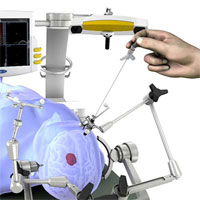
- Systems using the patient’s spatial attachment are not in a space limited by a frame, but in the space around the operating table. In this case, the movements of the instrument are monitored in the hands of the surgeon and in real time it is reported where it is located.
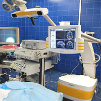
Frameless neuronavigation Vector Vision (BrainLab, Germany). We carried out the first stage of navigation — pre-operational planning, which consisted in the installation of virtual points of planned access for the preoperative construction of the optimal trajectory to the pathological focus, and, if necessary, made a three-dimensional construction of this focus. The access path was calculated in such a way as not to damage the functionally important zones.
Before the start of the operation (after the introduction of anesthesia) at a certain distance from the intervention area, a special navigation frame with a series of light-emitting diodes is rigidly fixed. To the Mayfield clamp was attached a kind of “antenna” — an active tracking infrared sensor.
Then a special pointer with a laser emitter of the surgeon delineates the surface of the patient’s head, using natural anatomical landmarks (brow ridges, lower edge of the eye socket, nose bridge, etc.). The system connects the three-dimensional image from its memory with the real position of the patient’s head. After registration, the navigation system delivers the accuracy of the patient’s head and virtual model on the display.
Under the control of the display data of the navigation installation in real time using an infrared probe, an economical cutaneous incision and craniotomy was planned, an optimal place of encephalotomy, a trajectory to the tumor and the border of the tumor were determined.
The surgeon can at any time, with an accuracy of
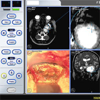
The position of the surgical instruments is monitored by monitoring the navigation system in three planes (axial, sagittal and coronary) at various stages of surgical intervention.
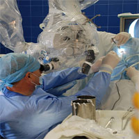
The reference frame is also fixed on the surgical microscope. Now any movements of the operating microscope are displayed on the navigation station monitor. On the monitor of the navigation station, the intraoperative pattern in the operating wound is combined from a microscope with a volumetric reconstruction of the brain, a pathological focus and vessels.
With the removal of meningiomas of the chiasmatic-selar region, the use of navigation and an operating microscope makes it possible to isolate and preserve the internal carotid and anterior cerebral arteries, prevent trauma of blood vessels in the arterial circle of the brain, and ensure the anatomical preservation of the stalk of the pituitary gland and optic nerves.
When the intracerebral neoplasms of the functionally important brain regions are removed with high accuracy, it is possible to identify the location of the motor and sensitive cortical areas, as well as the main speech centers (Broca and Wernicke), and their topographic relationship with the tumor. Three-dimensional (3D) reconstruction of cortical veins plays an important role in the visualization of large veins in the projection of volumetric education. These methods, with their combined use, make it possible to plan surgical access in such a way as to reduce the likelihood of damage to functionally important zones, cerebral vessels, to reduce the trauma of healthy brain tissue while striving for maximum tumor resection.
During the removal of the neoplasm, neuronavigation allows you to accurately orientate in the surgical site, optimize access to the tumor, taking into account the morphological and functional features of the paratamorous zone.
Today, navigation technologies have become widespread in practical neurosurgery. Frameless navigation based on preoperative CT and MRI allows planning surgical access, minimizing cutaneous incision, reducing the size of trephination, but it does not take into account the change in anatomy of the brain during surgical intervention. The reasons for the change in anatomy are, first of all, removal of tumor volume, brain edema, and loss of cerebrospinal fluid, which lead to brain displacement. To solve this problem, various methods of intraoperative imaging (CT, MRI, ultrasound diagnostics)
Frameless biopsy with the use of the neuronavigation system of intracerebral tumors of functionally important brain areas, diffuse lesions in the cerebral hemispheres, deep interhemispheric localization is an adequate alternative to the framework stereotaxis.


